
01 / ROI plane
Start with the regions.
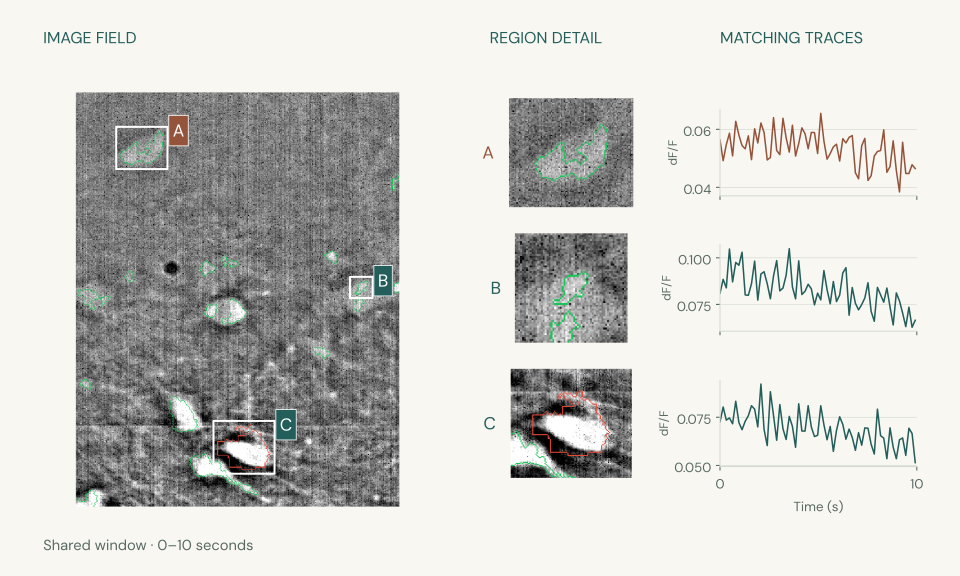
Identify regions in the image and keep each one connected to its trace.
- Field of view
- Region review
- Extracted traces
Research / Calcium imaging
We build calcium imaging workflows for region selection, trace extraction, and network exploration. Researchers can inspect the methods, parameters, and quality-control decisions behind the analysis.
Before the analysis
Retain the microscope and detector setup, indicator, frame timing, and spatial scale with each recording. These acquisition details help researchers assess region morphology and interpret fluorescence dynamics.
From the platform
Image analysis, event-level activity and network exploration, brought together around your research.
01 / ROI plane
Identify regions in the image and keep each one connected to its trace.
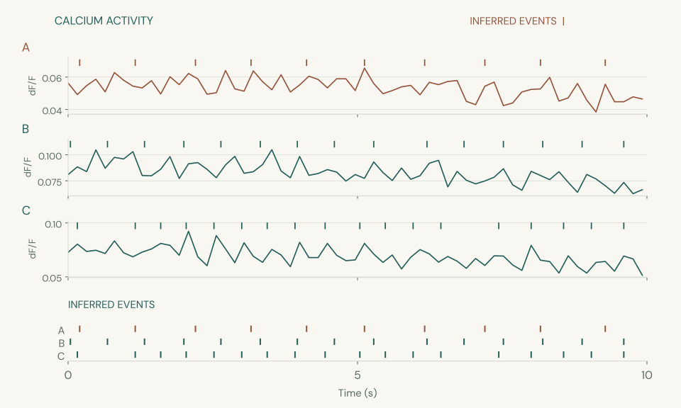
02 / Event plane
Connect fluorescence traces to inferred calcium events, with the timing available to explore.
03 / Network plane
Explore functional coactivity across regions and bring the wider pattern into view.
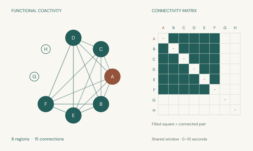
Adapted from an existing platform recording. Image pixels, trace samples, inferred event times and graph connections are retained. Labels and styling are adapted for presentation.
Network links show functional coactivity within the selected window. These examples demonstrate workflow capabilities; they do not establish biological findings or imply lab endorsement.
Methods & orchestration
Connect specialist imaging tools in a configured pipeline, from microscopy files to reviewable outputs. Retain processing parameters, intermediate artifacts, and QC decisions so your team can inspect each step.
Workflow
Stage dependencies, processing backends and expected outputs.
Parameter profile
ROI criteria, background correction, decay time and event thresholds.
Run record
Resolved settings, source references and the artifacts each stage produced.
Preflight → register → ingest
Check source metadata, establish dataset identity and materialize the movie while preserving the original input.
ND2, TIFF, and AVI are declared inputs. Retain timing metadata and frame selections with the recording; the workflow profile determines how the source is prepared.
Bounded native motion correction
Estimate frame shifts with phase correlation and carry the alignment record into downstream processing.
The native motion path records frame shifts, correction parameters and motion metrics. Its bounded entrypoint supports a separate motion run before post-motion processing.
Cellpose + CaImAn → candidate fusion
Bring morphology and CNMF-derived candidates together, retaining masks and the evidence behind each region.
The post-motion implementation invokes Cellpose and CaImAn CNMF initialization, then combines candidate masks with native morphology processing. Candidate generation is distinct from accepting a cell.
Mask-based extraction + ROI review
Connect raw fluorescence, background-corrected traces and ΔF/F to cell labels and merge decisions.
The v2 profile includes annular and shared-background correction. The reviewed path finalizes a snapshot of scientist decisions before re-extracting traces from reviewed masks; configured automated ROI review is also supported.
Reference OASIS → event analysis
Apply calcium deconvolution and evaluate candidate events alongside signal quality and trace evidence.
The reviewed downstream path uses reference OASIS AR(1), with decay time, baseline and event thresholds recorded in its settings. Inferred activity remains an estimate for scientific review.
Quality summary → package → archive
Package figures, tables, manifests, and run parameters together so researchers can trace the outputs to the processing that produced them.
Packaging collects analysis artifacts and provenance. The export stage records checksums and destination receipts; the configured output set determines what is delivered. Figure tooling includes Matplotlib for static exports.
A stage produces the inputs for the next. Profiles carry the method choices; run records carry what happened. A finalized ROI review can feed a downstream run that reuses the motion-corrected movie.
Tools within the workflow
Based on the current Calcium Scientific v2 workflow and its bounded processing paths. Steps are grouped for readability; enabled backends and delivered outputs depend on the run configuration.
Scientists in the workflow
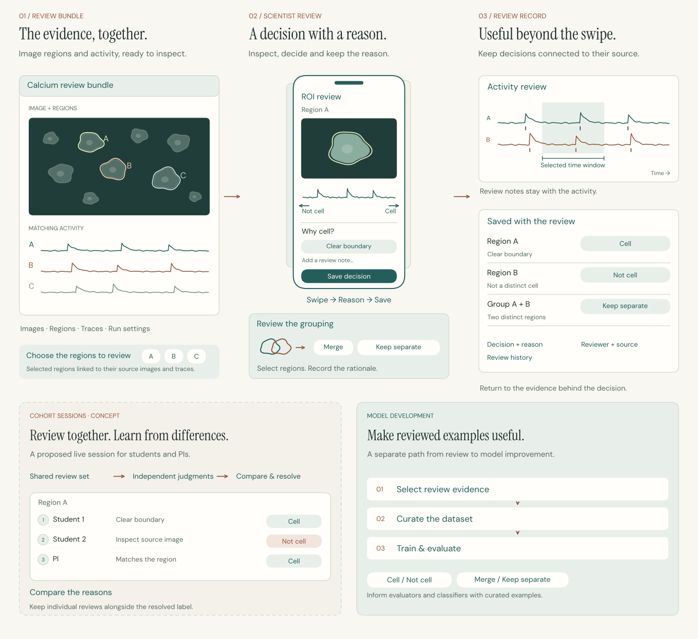
Review on desktop or mobile, keep the reasons with the evidence, and make each review useful for the next round of work.
Research operations
Carry the source movies, region sets, traces and review decisions forward with the method and settings used. Keep that record useful for the next experiment, method comparison or researcher joining the lab.
A conversation, grounded in your work
Tell us about the preparation, the current analysis, and where your lab needs support.